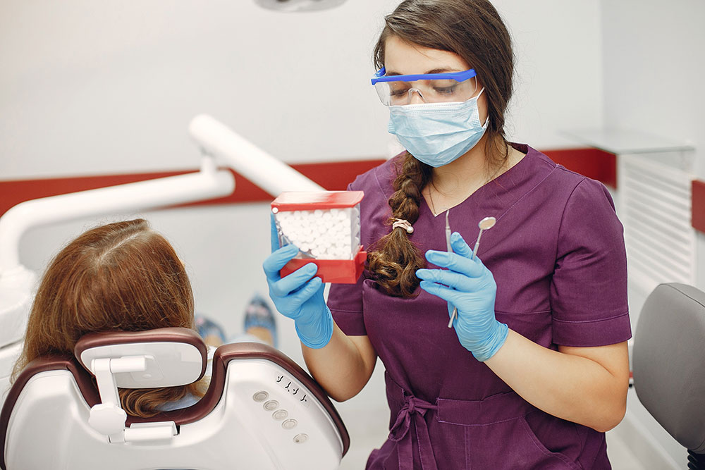
Dentistry has entered the digital age, allowing for greater accuracy, reduced discomfort, and enhanced patient communication. From the digitalization of impressions and the use of artificial intelligence in diagnostics to the incorporation of laser technology and 3D printing for dental prosthetics, the possibilities seem limitless. Dental technology has undergone a remarkable transformation in recent years, revolutionizing the way oral health is assessed, diagnosed, and treated. In this rapidly evolving field, cutting-edge innovations are changing the landscape of dentistry, providing patients with improved outcomes and experiences while empowering dental professionals to deliver more precise and efficient care.
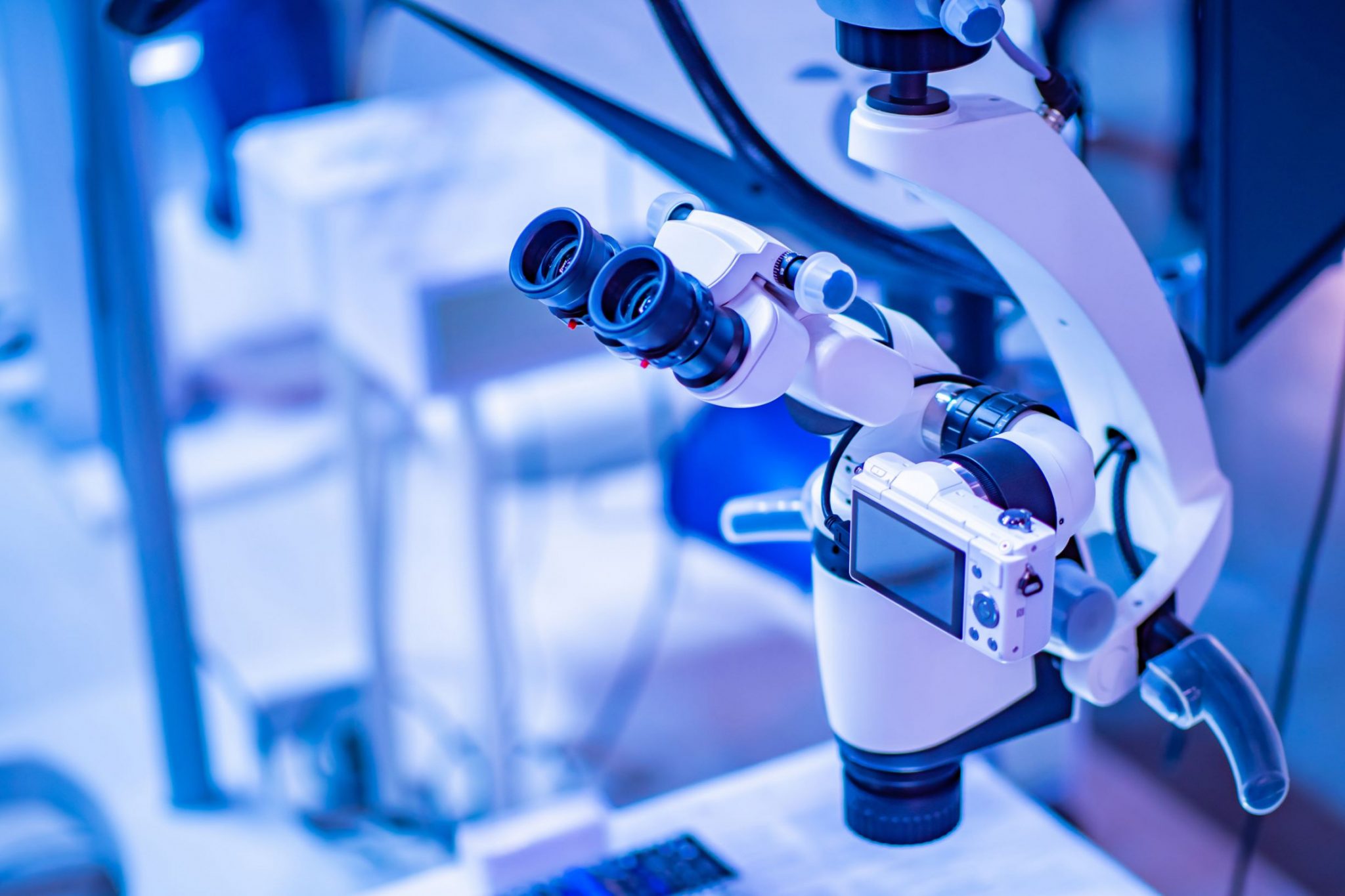
Dental microscope:
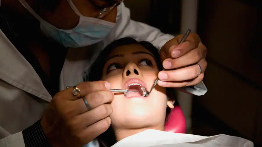
A dental microscope, also known as a dental operating microscope or dental microscope, is a specialized optical instrument used by dentists and oral surgeons to enhance their ability to diagnose and perform dental procedures with precision. These microscopes provide high levels of magnification and illumination, allowing dental professionals to see details within the oral cavity that are not visible to the naked eye.
Key features include:
Magnification: Dental
microscopes typically offer varying levels of
magnification, often ranging from 2x to 20x or more.
This magnification helps dentists examine teeth,
gums, and oral tissues in great detail.
Dental microscopes have numerous applications in dentistry, including:
Endodontics:
Dentists use
microscopes to perform root canal treatments
with precision, enabling them to locate and
treat even the smallest root canals.
Restorative
Dentistry:
Microscopes
help dentists achieve precise results in
procedures like dental fillings, crowns,
and veneers.
Oral
Surgery:
Oral
surgeons use dental microscopes for
procedures such as implant
placement, tooth extraction, and
tissue grafting.
Periodontics:
In
periodontal procedures, dental
microscopes aid in the accurate
assessment and treatment of gum
disease and other oral soft
tissue issues.
Orthodontics:
Dentists
and orthodontists use
microscopes for tasks like
bracket placement and the
precise adjustment of
orthodontic
appliances.
Dental microscopes have
significantly improved the
quality of dental care by
enabling dentists to
visualize and treat dental
issues with greater
accuracy. They also
contribute to patient
comfort and improved
treatment outcomes.
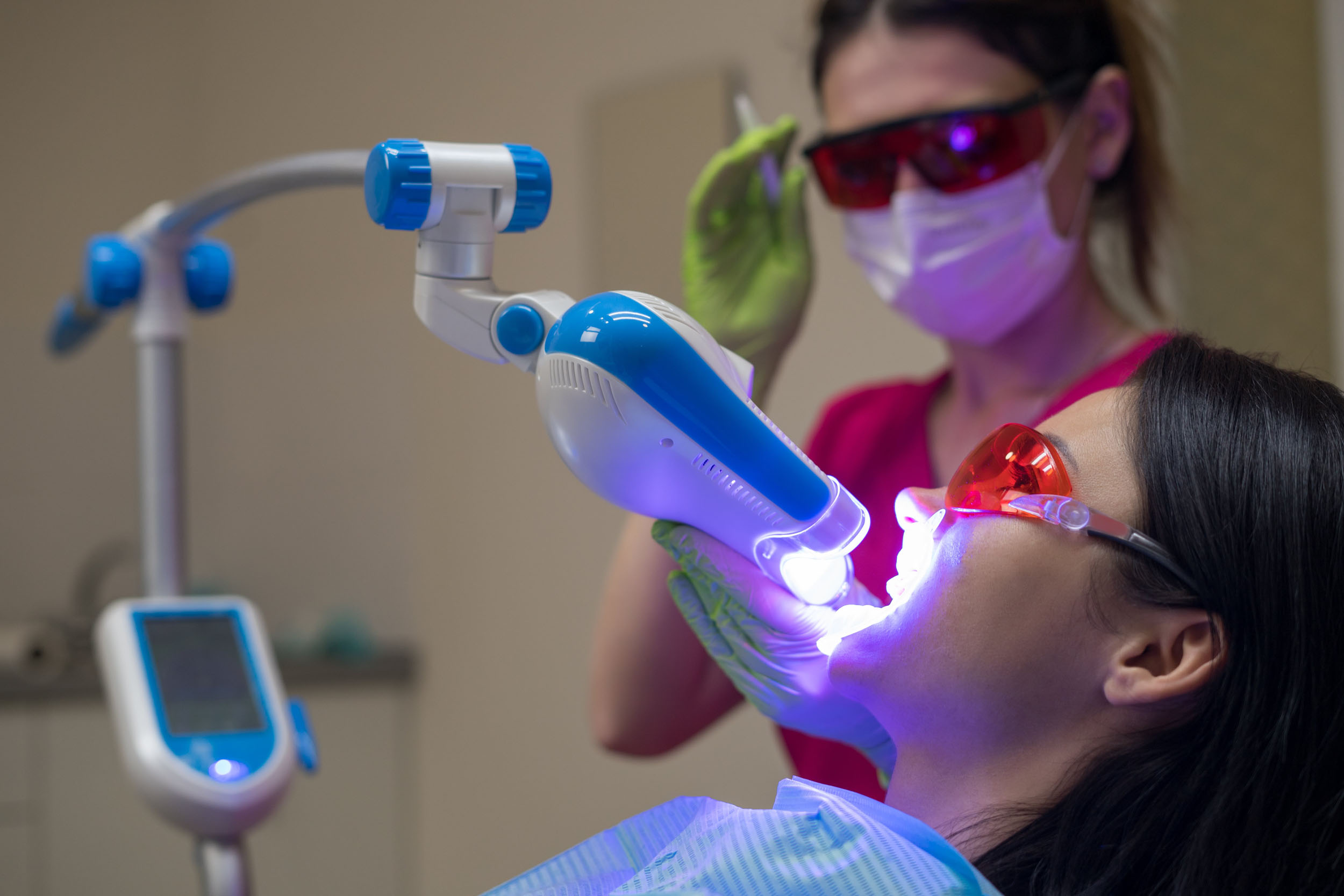
Laser technology continued to advance, enabling more precise and minimally invasive dental procedures. Dental lasers are specialized lasers used in various dental procedures to treat oral health issues. These lasers provide precise and minimally invasive alternatives to traditional dental tools like drills and scalpels.
Some common types of dental lasers include:
Soft
Tissue Lasers:
These lasers are used
for procedures involving the gums,
mucous membranes, and other soft
tissues in the oral cavity. They are
often employed for procedures like
gum contouring, removal of excess
tissue, and treatment of periodontal
(gum) disease.
Hard
Tissue Lasers:
Hard tissue lasers are
designed for dental work involving
teeth and bones. They are commonly
used for procedures like cavity
preparation, removal of tooth decay,
and enamel etching for bonding
procedures.
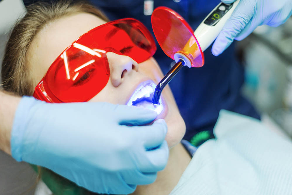
Diode
Lasers:
Diode
lasers are versatile and can be used
for a variety of dental
applications, including soft tissue
procedures like gum reshaping and
hard tissue procedures like teeth
whitening.
Dental lasers offer several
advantages, including reduced
discomfort for patients, faster
healing times, and often the
elimination of the need for
anesthesia. They also minimize the
risk of infection and can be more
precise than traditional tools.
Dentists use specific wavelengths
and power settings tailored to the
procedure and the patient's needs.
However, not all dental procedures
can be performed with lasers, and
their use depends on the dentist's
expertise and the patient's specific
case.
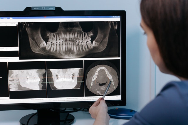
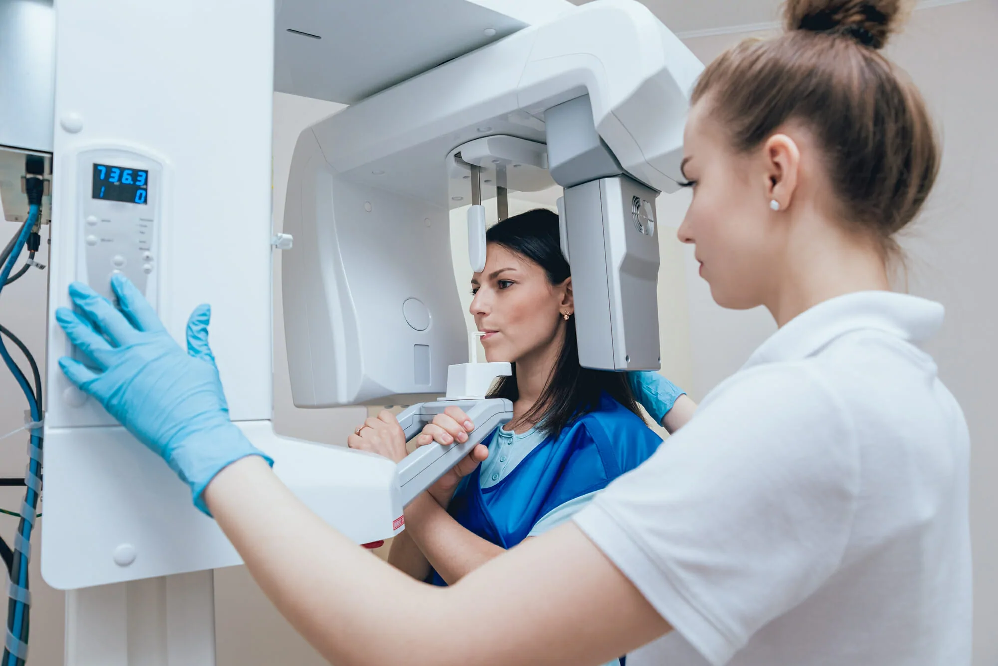
CONE
BEAM CT:
Cone
Beam Computed Tomography
provided 3D imaging for more
accurate diagnosis and treatment
planning. Dental Cone Beam
Computed Tomography (CBCT) is a
specialized type of medical
imaging equipment used in
dentistry to capture detailed 3D
images of the oral and
maxillofacial region, including
the teeth, jaws,
temporomandibular joints (TMJ),
and surrounding structures. Here
are some key points about dental
CBCT:
3D
Imaging:
Unlike traditional dental
X-rays that provide 2D images,
CBCT technology offers a
three-dimensional view of the
dental and craniofacial
structures. This allows for more
accurate diagnosis and treatment
planning.
Applications:
Dental CBCT is used for a
wide range of purposes in
dentistry, including implant
planning, oral surgery,
orthodontic assessment,
endodontic evaluation, and the
diagnosis of various dental and
facial conditions.
Low
Radiation:
While CBCT involves radiation
exposure, the doses used in
dental CBCT are generally lower
than those of medical CT scans,
making it a safer option for
dental imaging.
Precise
Measurements:
CBCT images
provide precise measurements and
allow for the assessment of bone
density and quality, which is
crucial for dental implant
planning.
Treatment
Planning:
Dentists and oral
surgeons use CBCT images to plan
complex dental procedures such
as implant placement, root canal
therapy, and orthognathic
surgery. It helps in determining
the optimal location and angle
for dental implants.
TMJ
Evaluation:
CBCT is valuable
for evaluating temporomandibular
joint disorders (TMJ) and
assessing the relationship
between the jaw joint and
surrounding structures.
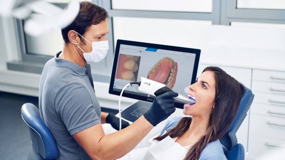
INTRAORAL
CAMERAS:
High-resolution
intraoral cameras were
being used to provide
real-time images of
patients' mouths for
better diagnosis and
patient education.An
intraoral dental scanner
is a digital imaging
device used by dentists
to capture detailed 3D
images of a patient's
oral cavity, including
teeth and gums. It
replaces traditional
dental impressions,
which can be
uncomfortable for
patients. These scanners
use technologies like
laser or structured
light to create highly
accurate digital models,
making it easier for
dentists to plan and
execute procedures such
as crowns, bridges, and
braces. Intraoral
scanners improve patient
comfort and save time
compared to traditional
methods.
They have become an
essential tool in modern
dentistry.
Intraoral scanners are
advanced dental devices
used to create highly
accurate digital
impressions of a
patient's teeth and oral
structures. These
devices have become
increasingly popular in
modern dentistry due to
their numerous
advantages over
traditional
impression-taking
methods, such as using
molds and trays filled
with impression
material. Here are their
uses and advantages:
Orthodontics:
Intraoral
scanners are used to
capture digital
impressions of a
patient's teeth, which
can then be used to plan
and monitor orthodontic
treatments, such as
braces or aligners.
Restorative
Dentistry:
Dentists
use intraoral
scanners to create
digital impressions
of teeth for the
fabrication of
dental crowns,
bridges, inlays,
onlays, and veneers.
These impressions
are sent to a dental
laboratory for
precise restoration
fabrication.
Implant
Planning:
Intraoral scans
aid in planning
dental implant
procedures by
providing
detailed images
of the patient's
oral anatomy.
This ensures the
precise
placement of
implants.
Prosthodontics:
For
the creation
of removable
or fixed
partial or
complete
dentures,
intraoral
scans
provide
accurate
impressions
to design
and
fabricate
prosthetic
devices.
Periodontics:
Intraoral
scanning
can help
periodontists
assess
the
condition
of the
gums and
teeth,
track
changes
in gum
health,
and plan
gum
disease
treatments.
Advantages:
Precision:
Intraoral
scanners
offer
high
precision
and
accuracy,
reducing
the
chances
of
errors
compared
to
traditional
impression
methods.
Patient
Comfort:
Patients
often
find
intraoral
scanning
more
comfortable
than
traditional
impressions,
as
it
eliminates
the
need
for
messy
impression
materials
and
trays
that
can
induce
gagging.
Speed:
Intraoral
scanning
is
faster,
significantly
reducing
chair
time
for
patients.
This
can
lead
to
improved
patient
satisfaction
and
higher
practice
efficiency.
Digital
Records:
Digital
impressions
are
stored
electronically,
making
them
easy
to
access,
store,
and
share
with
other
healthcare
providers
or
dental
laboratories.
Enhanced
Communication:
Dentists
can
visually
share
digital
impressions
with
patients,
helping
them
better
understand
their
treatment
needs
and
options.
Reduced
Material
Waste:
Traditional
impressions
require
disposable
materials
like
trays
and
impression
compound,
contributing
to
waste.
Intraoral
scanning
is
more
environmentally
friendly.
Immediate
Feedback:
Dentists
can
immediately
assess
the
quality
of
digital
impressions
and
make
any
necessary
adjustments
during
the
scanning
process.
Better
Clinical
Outcomes:
The
accuracy
of
intraoral
scans
can
lead
to
better-fitting
restorations
and
orthodontic
appliances,
ultimately
improving
clinical
outcomes.
Remote
Consultations:
Digital
impressions
can
be
sent
electronically
to
dental
laboratories
or
specialists
for
remote
consultations,
expediting
treatment
planning.
Long-term
Cost
Savings:
While
the
initial
investment
in
an
intraoral
scanner
can
be
significant,
it
can
lead
to
cost
savings
over
time
due
to
reduced
material
costs
and
increased
efficiency.
In
summary,
intraoral
scanners
are
versatile
tools
that
benefit
both
dental
practitioners
and
patients
by
providing
precise
digital
impressions
for
a
wide
range
of
dental
procedures.
Their
advantages
include
increased
accuracy,
improved
patient
comfort,
time
savings,
and
enhanced
communication.